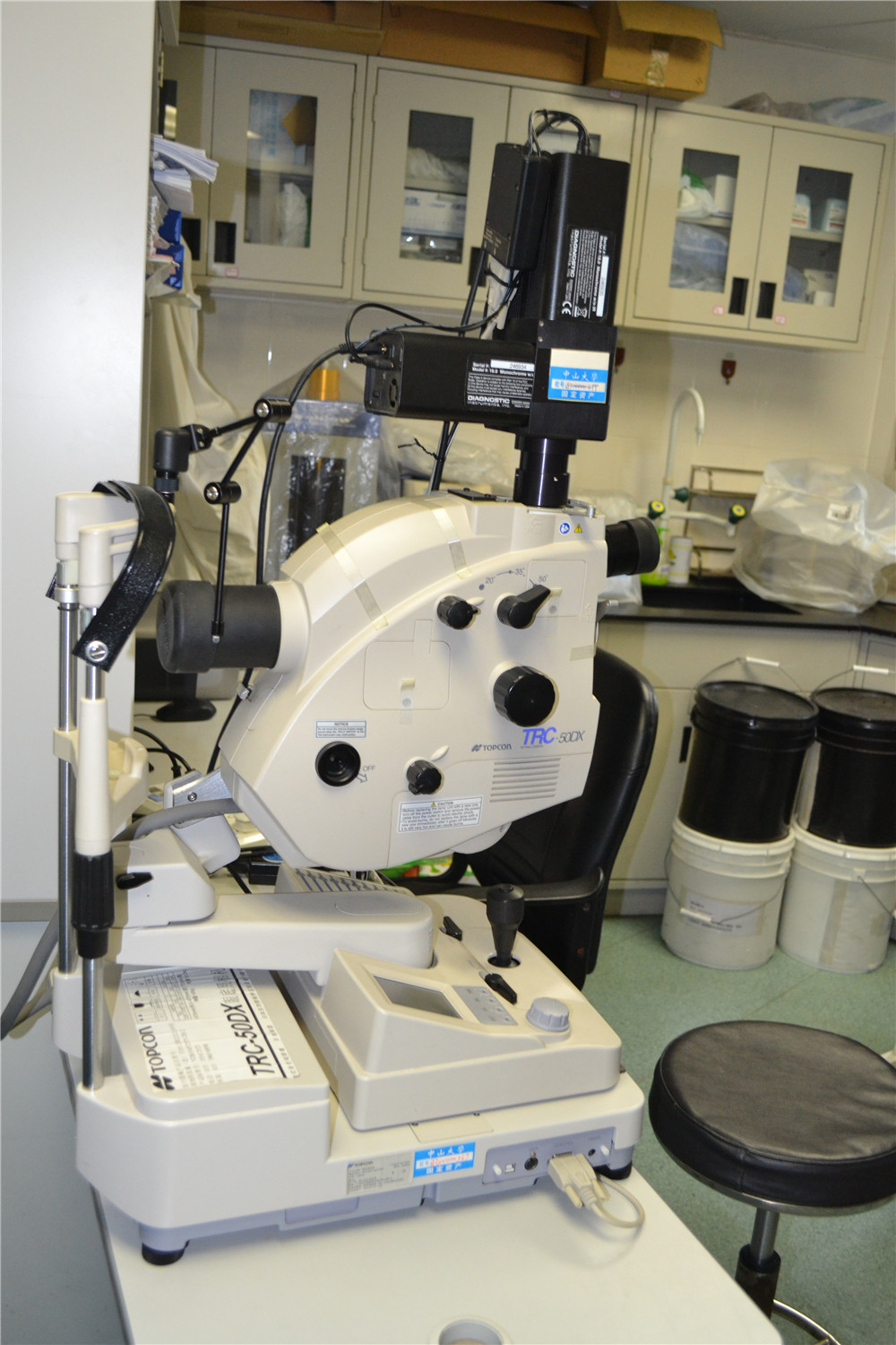
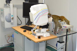
Although different retinal oximeter systems have slightly different constructions, the basic retinal oximeter consists of a fundus camera with an attached image splitter. The image splitter contains mirrors that split the beam from the camera into smaller beams based on wavelengths of light, as well as filters that further filter the beam. The apparatus also includes a digital camera to record the fundus images. Analysis of the images has been shown to be most effective with comparison of the images at two distinct wavelengths, at about 600 nm (sensitive to oxyhemoglobin) and about 570 nm (not sensitive to oxyhemoglobin). Computer software detects retinal vessels and uses relative light intensities to calculate relative vessel oxygenation. Multivariate regression analyses of studies on normative values in healthy subjects has shown no significant associations between retinal oxygen saturation (SO2) or vessel width and iris pigmentation, ethnicity, gender, and smoking status. Aging, however, is a factor that has been shown to decrease retinal venous (SvO2) and arterial oxygen saturation (SaO2) and affects most retinal SO2 parameters and therefore must be accounted for in interpreting and comparing measurements.
相关文章
-
 2023-03-29
 - 2023-03-29
- 2023-03-29
- 2023-03-29